Screening Procedures
General Overview
Radiographs submitted to the OFA should follow the American Veterinary Medical Association recommendations for positioning. This view is accepted world wide for detection and assessment of hip joint irregularities and secondary arthritic hip joint changes. To obtain this view, the animal must be placed on its back in dorsal recumbency with the rear limbs extended and parallel to each other. The knees (stifles) are rotated internally and the pelvis is symmetric. Chemical restraint (anesthesia) to the point of relaxation is recommended. For elbows, the animal is placed on its side and the respective elbow is placed in an extreme flexed position.
The radiograph must be permanently identified with the animal’s registration number or name, the date the radiograph was taken, and the veterinarian’s name or hospital name. If this required information is illegible or missing, the OFA cannot accept the film for registration purposes. Both the owner and vet should complete and sign their respective sections of the OFA application. It is important to record on the OFA application the animal’s tattoo or microchip number in order for the OFA to submit results to the AKC. Sire and dam information should also be present.
Radiography of females in estrus or pregnant should be avoided due to possible increased joint laxity (subluxation) from hormonal variations. OFA recommends radiographs be taken one month after weaning pups and one month before or after a heat cycle. Physical inactivity because of illness, weather, or the owner’s management practices may also result in some degree of joint laxity. The OFA recommends evaluation when the dog is in good physical condition.
Chemical restraint (anesthesia) is not required by OFA but chemical restraint to the point of muscle relaxation is recommended. With chemical restraint, optimum patient positioning is easier with minimal repeat radiographs (less radiation exposure) and a truer representation of the hip status is obtained.
For large and giant breed dogs, 14″ x 17″ film size is recommended. Small film sizes can be used for smaller breeds if the area between the sacrum and the stifles can be included.
If a copy is necessary, ask your veterinarian to insert 2 films in the cassette prior to making the exposure. This will require about a 15% increase in the kVp to make an exact duplicate of the radiograph sent to OFA. Films may be returned if a $5.00 fee and request for return are both included at the time of submission.
Good contrast is desirable (high mAs, low kVp). Grid techniques are recommended for all large dogs.
Radiation Safety: Proper collimation and protection of attendants is the responsibility of the veterinarian. Gonadal shielding is recommended for male dogs.
OFA Handling Procedures
When a radiograph arrives at the OFA, the information on the radiograph is checked against the information on the application. The age of the dog is calculated, and the submitted fee is recorded. The board-certified veterinary radiologist on staff at the OFA screens the radiographs for diagnostic quality. If it is not suitable for diagnostic quality (poor positioning, too light, too dark or image blurring from motion), it is returned to the referring veterinarian with a written request that it be repeated. An application number is assigned.
Radiographs of animals 24 months of age or older are independently evaluated by three randomly selected, board-certified veterinary radiologists from a pool of 20 to 25 consulting radiologists throughout the USA in private practice and academia. Each radiologist evaluates the animal’s hip status considering the breed, sex, and age. There are approximately 9 different anatomic areas of the hip that are evaluated.
- Craniolateral acetabular rim
- Cranial acetabular margin
- Femoral head (hip ball)
- Fovea capitis (normal flattened area on hip ball)
- Acetabular notch
- Caudal acetabular rim
- Dorsal acetabular margin
- Junction of femoral head and neck
- Trochanteric fossa
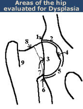
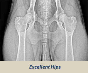
The radiologist is concerned with deviations in these structures from the breed normal. Congruency and confluence of the hip joint (degree of fit) are also considered which dictate the conformation differences within normal when there is an absence of radiographic findings consistent with HD. The radiologist will grade the hips with one of seven different physical (phenotypic) hip conformations: normal which includes excellent, good, or fair classifications, borderline or dysplastic which includes mild, moderate, or severe classifications. See more on Hip Grades.
Submitting Radiograph Screenings
For more details on how veterinary clinics should submit radiograph screenings of hips and elbows, with options via email and mail, see the Veterinary Submissions Procedures.